Slipped Disc X Ray Or Mri

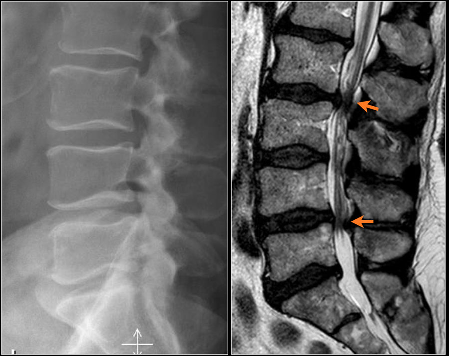
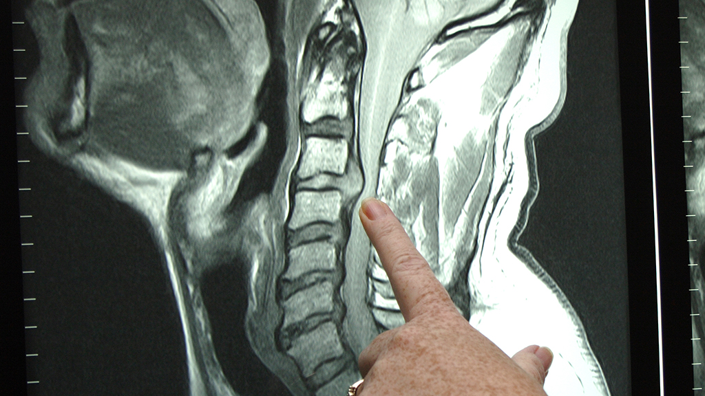
A traditional x ray shows bony ridges common to spinal injuries and disorders such as cervical spondylosis but seldom shows soft tissue damage.
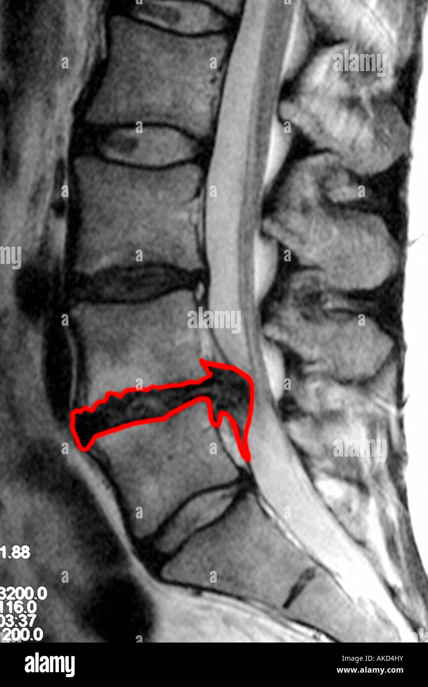
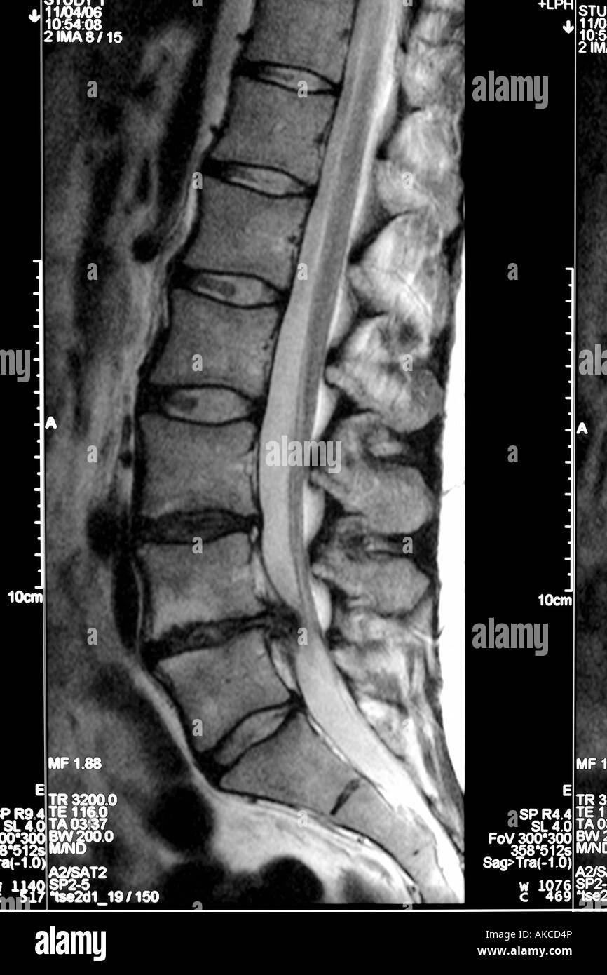
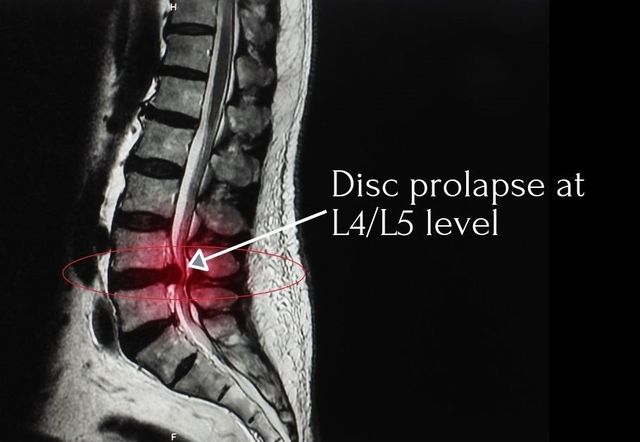
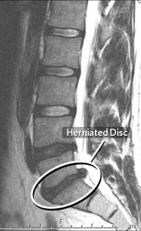
Slipped disc x ray or mri. By comparison a herniated disc on an mri. Directly to the online appointment arrangement. You can find me in. Your doctor may recommend an x ray to look at the vertebrae surrounding a herniated disc.
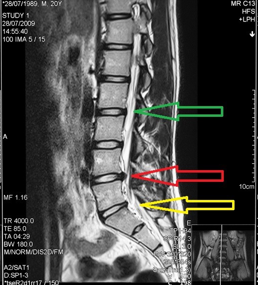
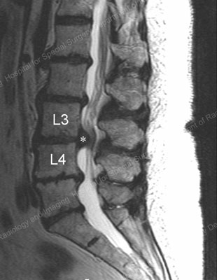
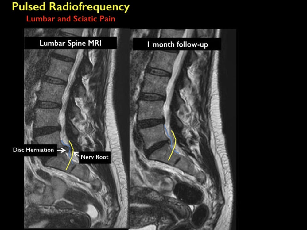
Plain x rays don t detect herniated disks but they can rule out other causes of back pain such as an infection tumor spinal alignment issues or a broken bone. A herniated disc is a disease that is characterized by the protrusion of parts of the disc into the spinal canal. Often if a disc slips out of place the space between vertebrae may shrink or the vertebrae may become unstable without the disc to act as a cushion. A ct scan may show evidence of a ruptured disc.
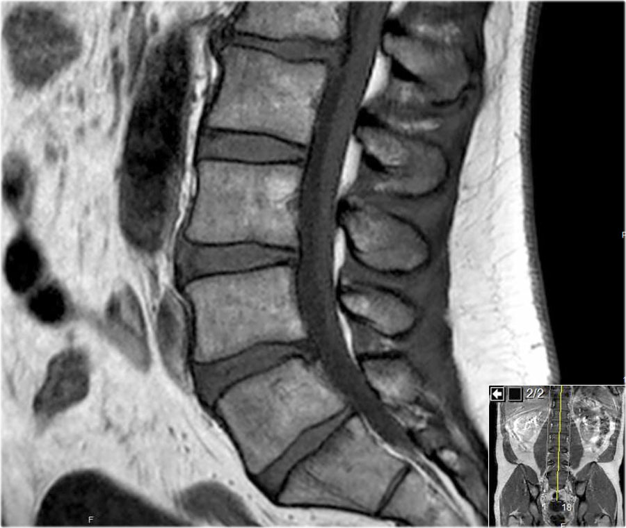
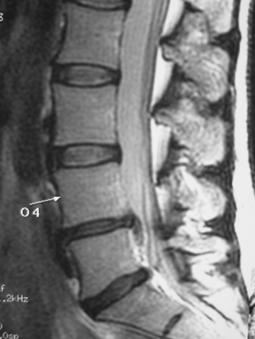
While a standard x ray can t show if you have a herniated disk it can show your doctor the outline of your spine and rule out whether your pain is caused by something else such as a. A ct scanner takes a series of x rays from different directions and then combines them to create cross sectional images of your spinal column and the structures around it. Magnetic resonance imaging mri scans are the best tools for diagnosing a slipped disc. X rays use high energy beams of light to create detailed images of the spine.
X ray ultrasound mri etc be assessed.
